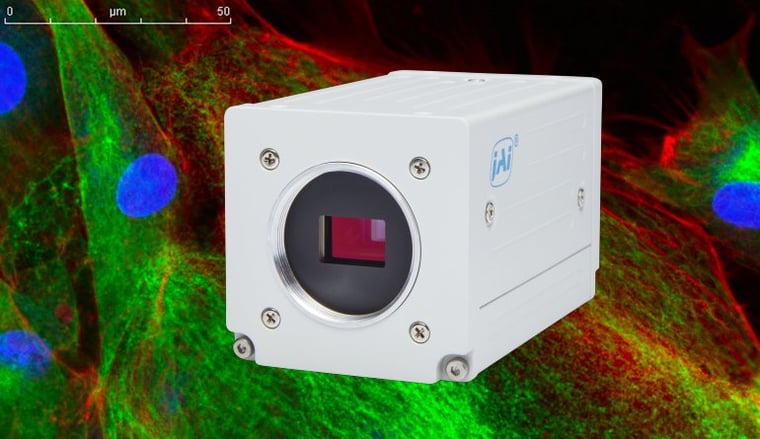
Color imaging is being used in more and more life sciences applications these days. In the field of fluorescence microscopy, the use of color is particularly important as it enables different proteins or other macromolecules to be mapped and studied simultaneously by attaching fluorophores that emit at different wavelengths. In some cases, this analysis is still done by assigning multiple filtered grayscale images to different color channels in a graphics software program. But the use of modern color cameras can capture these multi-color fluorescence images in a single step.
Autofluorescence
One obstacle to capturing good multi-color fluorescence images, especially of living tissues, is autofluorescence. Autofluorescence is a natural fluorescence that occurs in most living tissues, largely due to biological substances such as mitochondria, lysosomes, flavins, porphyrins and chlorophyll. The spectral response of autoflourescence is generally broadband and dominant in the green wavelengths.
In microscopic techniques that use fluorescence or phosphorescence to study the properties of organic and inorganic materials, this green autofluorescence interferes with the detection of fluorescent contrast agents, causing structures other than those of interest to become visible and generating a high background signal that reduces the overall contrast and clarity in such images. The problem is further complicated when the cell culture media consists of cellular extracts which can also emit autoflourescence.
Pushing the boundaries
One way to minimize the effect of autofluorescence is to spread the colors in the image farther apart in spectral terms. The effect of autoflourescence degrades naturally at wavelengths above 700 nm which makes it advantageous to use a red fluorescent dye that emits wavelength across the visible/NIR boundary, this way it receives less interference from the autofluorescence “noise” in the image.
There are several fluorophores such as ATTO594, Allophycocyanin, Cy5TM, ATTO 647N, DyLightTM 649, ATTO 655, Cy5.5TM, DyLightTM 680, DyLightTM 755, DyLightTM 800, ATTO 700 which absorb light between 500 nm – 700 nm and emit flourescence between 600 nm and 1000 nm. This is a spectral range which can be called “red-to-extended-red (NIR)” in conventional camera technology terms.
Those with emission plots starting below 700 nm and continuing into the NIR range are useful for generating a more distinctive red response in multi-color images. Those with emission plots starting above 700 nm and ending below 1000 nm are more accurately classified as NIR flourescence dyes (known as the NIR-I spectrum in medical imaging terms). These have several advantages over conventional visible dyes such as deeper penetration of NIR energy and much better image contrast at increased tissue thicknesses.
Need some guidance in creating microscopy systems with advanced color capabilities?Download our Tech Guide: Advanced Color Microscopy Solutions, and learn how various camera capabilities can be utilized to meet your application requirements.
Camera considerations
For builders of microscopy-based imaging systems, the use of color cameras with these red-to-extended-red fluorophores is severely limited unless the color camera has support for a wider response in the red/NIR range. Most single-sensor color cameras used in microscopy are equipped with an NIR-cut filter that provides a spectral cut-off around 700 nm or even lower. This is because the cameras use a pattern of color filters over the pixels and a process of interpolation to estimate each pixel’s color based on the value of surrounding pixels. If the NIR filter is removed and NIR light is allowed to affect the intensity values of the red pixels in the image, it distorts the values of all the other pixels during the interpolation process. As a result, these cameras are limited to fluorophores that stay completely within the confines of the visible spectrum.
Three-sensor, prism-based color cameras provide a possible solution for applications needing extended red sensitivity for life sciences applications. Removing the normal NIR-cut filter from a prism camera extends the sensitivity of the red channel to roughly 1000 nm, enabling it to boost the response from fluorophores that straddle the visible-NIR boundary or fall entirely in the NIR-I range. But because the R-G-B channels are independent color planes with no interpolation required, the effect of this extended red sensitivity on the other color channels is minimized.
JAI offers several three-sensor prism cameras specially designed to serve the life sciences microscopy market, including models with no NIR-cut filter on the red channel for extended sensitivity in the red/NIR waveband. For questions about extended red sensitivity or for additional information about JAI’s life sciences cameras, please contact JAI.
