
The field of medical imaging is evolving rapidly. With the highly potent combination of advanced imaging and high-tech image analysis, we are now able to visualize and study human physiology in high detail. Over the last few decades, imaging methods have evolved from conventional microscopes to cutting edge technologies. Given the imaging trends from the past and the medical challenges we face today, we can be certain that new methods of detection and identification will not only play a significant role in the healthcare industry but also in the future practice of medicine.
One of the cutting-edge technologies is multispectral imaging (MSI). This method involves using a limited number of spectral bands targeted at solving challenges in very specific applications.
From visible to multispectral imaging in life sciences applications:
Some imaging techniques such as microscopy or endoscopy are already well established. Modern RGB-endoscopes, for example, use sensors mounted at the distal tip of the endoscope to deliver high resolution images while having an extremely small diameter. Some are equipped with narrow band filtering techniques, allowing even more specific tissue inspection. The images generated by RGB-endoscopes are however limited to the visible spectrum and can only image the outermost tissue layer due to the inability of visible light to penetrate through the surface of the skin.
On the other hand, multispectral cameras capture information for multiple discretely-positioned spectral bands, including bands outside the visible region. When examining human tissue, the near infrared (NIR) wavelengths penetrate deeper into the tissue. This allows the physician to extract intrinsic properties and structures of specific tissues which are not visible to a human eye.
Intrinsic tissue properties, such as molecular composition, can be helpful for the physician to diagnose and monitor a patient. The physician would usually have to take a biopsy, which comes with several disadvantages as it is invasive, time consuming, unpleasant for the patient, and results in loss of tissue. Using MSI cameras, tissue properties can be visualized in real time without the need for a biopsy.
As incident light propagates in human tissue, it gets scattered and absorbed. Both are tissue dependent processes. Absorbed light is either transformed into heat or radiated during a process called photoluminescence. Fluorescence, which is one of the forms of photoluminescence, is of specific importance during this process. Depending on the molecular structure, tissues can have different physical footprints which can be characterized based on its fluorescence spectrum. For example, the fluorescence emitted by healthy tissue is different than from scarred or a damaged tissue.
Malignant or benign lesions caused by a tumor result in pathological changes in the tissue and alter its scattering, absorption, and fluorescence properties. Increased metabolic activity of tumor cells can increase the hemoglobin concentration.
Hemoglobin is a chromophore which increases the interaction with light. Furthermore, alterations in the metabolic pathway and molecular structure would change the fluorescence properties of the tissue.
Multispectral imaging in hematopathology:
Multispectral imaging can also be used in hematopathology. Relevant quantitative morphological and molecular information of the blood sample is needed by the pathologist to carry out the diagnosis. Current techniques such as brightfield chromogenic imaging is limited to the visible spectrum. Multispectral imaging enables a convenient and robust way to conduct blood sample examination using not only visible wavelengths but also beyond.
Multispectral cameras for surgical guidance systems:
Surgical guidance is another promising application of multispectral cameras used in healthcare. The removal of a tumor is complicated and often requires critical surgery. The surgeon needs to be able to see and identify the tumor cells and accurately measure the margins to the surrounding healthy tissue. Removing too much tissue can harm the patient, removing not enough tissue will allow the tumor to grow back. Judgement is difficult and risky, even among specialists.
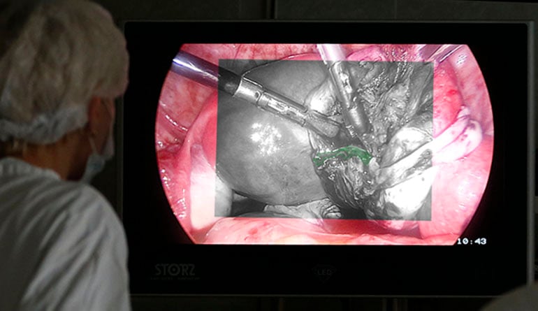
Multispectral imaging can add tremendous value in medical applications, especially in surgical tasks. Combining color imaging with NIR bands can help to locate and distinguish between tumors and surrounding tissues. In a real surgical situation, ICG might be injected into blood vessels, tissue or lymphatic vessels. With real-time video images overlaid on the visible image, surgeons could use fluorescence to locate tumors/glands for removal, highlight key vessels, and/or monitor blood flow while operating.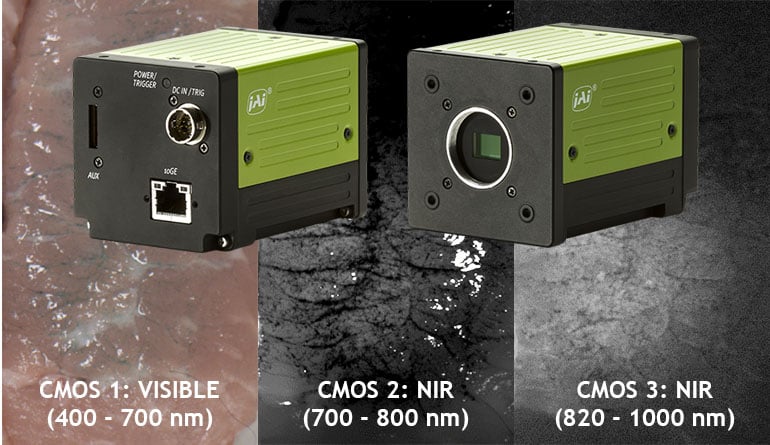
JAI's 3-CMOS multispectral prism cameras provide simultaneous images of three different spectral bands in a single camera - a visible color channel from 400-700 nm, a near infrared (NIR) channel from 700-800 nm, and a second NIR channel from 820-1000 nm. This makes it possible to simultaneously detect or inspect visible elements, as well as analyze materials or defects using their spectral characteristics within a single NIR band or when combined across both NIR bands.
Using near-infrared fluorescence in combination with visible imaging, the tumor can be highlighted in real time providing very high accuracy of the tumor location and its surroundings. Fusing the information from all the spectral bands provides the doctor with an augmented view during the surgery. Furthermore, the blood flow and its oxygen distribution can be visualized in real time.
Imaging in digital pathology and disease detection:
Multispectral imaging has great potential in digital pathology, diagnosis and disease detection, patient monitoring, surgical guidance and other healthcare applications. However, there are technical limitations of the camera systems that are used in daily patient care and diagnosis. The spectral imaging devices used in medical research are often very sophisticated (i.e., hyperspectral), complicated to use, and not commercially viable especially in developing and disease-ridden countries. There is a need for simpler and more affordable imaging systems which are equally effective and can solve specific challenges pertaining to medical imaging.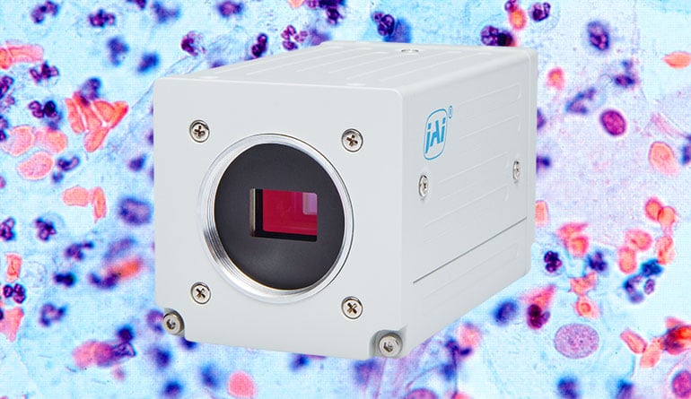
In recent years the field of digital pathology (where trained pathologists look at human tissue whole slides and cell samples under a microscope) has become more and more digitized and
automated, using high quality/high resolution color and multispectral imaging techniques, and image analysis software.
Improving imaging in life sciences applications with JAI prism-based multi-sensor cameras:
Prism-based multi-sensor cameras provide high resolution, high frame rates, and simultaneous imaging of visible and non-visible wavebands through a single optical axis. Prism-based multispectral cameras have high shock and vibration specifications to support the need for high robustness, which is vital for imaging devices used in medical applications. Furthermore, prism-based multispectral cameras can be combined with endoscopes, microscopes and other medical imaging devices to get the best of both worlds.
Learn more about JAI's multispectral cameras:
Download Tech Guide: Multispectral imaging:
Download this FREE tech guide about multispectral imaging for medical and industrial machine vision systems.
Watch webcast: Using prism-based cameras for multispectral applications:
If you are a vision system designer with a project that needs multispectral imaging capability, this free webinar can help you decide whether a prism-based multispectral camera is the right approach. There are several different types of multispectral cameras currently available and the one you select can have a major impact on your project’s success.
Contact JAI for help:
Let us help you to find the perfect camera to meet your application requirements in your life sciences application.