
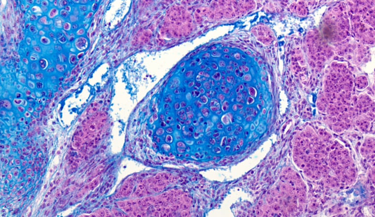
Prism color microscopy image courtesy of Digital Imaging Systems Ltd., UK
Two types of noise
For brightfield microscopy, the most important type of noise is random noise, sometimes referred to as “shot noise.” Random noise is the random variation of brightness or color information in images caused by fluctuations in the arrival of photons at the image sensor. These small, random intensity fluctuations interfere with the fine details in an image, and vary over time (they are different from frame to frame). While the best way to deal with random noise is to try to minimize its occurrence (see the section below on color balancing for brightfield microscopy), you may be able to reduce the effect of random noise by averaging several consecutive images or frames. Of course, this requires that the specimen does not move, nor is there any motion or vibration in the equipment or its surroundings. The degree of noise reduction achieved by averaging is proportional to the square root of the number of images. This means that in order to reduce the noise by half, you would need to average four images; to reduce it to one fourth of the original, you would need to average 16 images; and so on. Unfortunately, frame averaging tends to soften the image (i.e., some blurring with loss of sharpness), which may be just as unacceptable as the noise in the original image. This is why methods that minimize the amount of noise generated are so important.
The other main noise source is read noise. Read noise is created within the camera electronics during the readout process as the electrons are subjected to the analog-to-digital conversion, amplification, and processing steps that enable an image to be produced. It is often referred to as “dark noise” because it is best observed by removing all light from the image and noting the pixel-to-pixel variations that occur strictly based on the sensor and camera’s readout circuits. This type of noise becomes particularly apparent when using long exposure times (more relevant to fluorescence microscopy than brightfield microscopy). The effect also gets accelerated at higher temperatures, which is why some microscopy cameras resort to expensive “cooling” techniques to maintain the cleanest possible images for light-limited situations.
Read noise is often described as a combination of this dark noise with something called “fixed pattern noise.” Like dark noise, fixed pattern noise is caused by variations in pixel electronics that produce a non-random pattern of intensity variations in the image. Also like dark noise, it is most visible under low-light conditions.
How gain affects noise
The signal level (brightness) of a digital image can be increased by applying “gain” to the image. Gain is simply a term for electronic amplification. However, when you “turn up the amplifier” on a digital signal, you will also be amplifying any noise (random or read noise) that may be contained within the image. In most brightfield microscopy applications, it will be the random noise component that will be most significant. Applying gain to the image increases the variance of the random noise, making the “static” in the image more and more noticeable and interfering with image analysis applications. For this reason, it is important to avoid applying significant amounts of gain if at all possible.
Need some guidance in creating microscopy systems with advanced color capabilities?Download our Tech Guide: Advanced Color Microscopy Solutions, and learn how various camera capabilities can be utilized to meet your application requirements.
Color balancing for brightfield microscopy
In brightfield microscopy, the color temperature of the light source is an important factor for image capture and analysis. As light sources have a characteristic radiation spectrum, the color temperature heavily influences the color of objects in the image. Different color temperatures produce backgrounds with slightly different hues and alters the color of cells and other objects in the image. In order to perform analyses where a specific color may have a special significance, it is important to properly balance the color response of the camera against the light source’s color characteristics.
In most cameras, color balancing (also called white balancing) is done by placing a white target in the field of view and applying gain to two of the three color channels (red, green, blue) in order to get the same response from all three channels. For convenience, some cameras also offer color temperature “presets” that allows you to simply pick the preset that matches the color temperature of your light source. In both cases, however, this process works by adding gain to two of the color channels, thus raising the noise level in the image.
While prism-based cameras, such as those in JAI’s Apex Series, offer both gain-based white balancing and color temperature presets, their three-sensor design also provides a third method for achieving white balance. Each sensor can have its shutter speed set independently from the other two sensors. Thus, white balancing can be achieved by proportionally adjusting the exposure times of each sensor to match the color temperature of the light source. This can require lengthening the exposure time of the two channels with the lowest response, or shortening the exposure time of the two channels with the highest response. In either case, no additional gain needs to be applied to the image, which keeps noise at a minimum level.
For applications requiring clean, low-noise images, without resorting to expensive “cooled” cameras, prism-based technology may provide an attractive alternative.
For more information, on low-noise cameras for brightfield microscopy, please contact JAI.