
JAI has announced a series of new camera models and software integrations which provide advanced color imaging capabilities to builders and users of microscopy-based imaging systems. The announcement includes six new models in JAI’s Apex Series of high performance 3-CMOS prism color cameras, as well as full integration with two of the most popular microscopy software solutions – Image-Pro® from Media Cybernetics and the µManager open source software package.
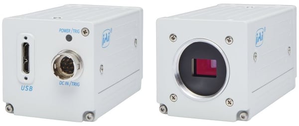
Three of the new JAI camera models are variations of the AP-3200T-USB, a 3.2-megapixel, 38.3 fps, 3-CMOS prism color camera. The other three models are variations on the AP-1600T-USB, a 1.6-megapixel 3-CMOS camera offering higher frame rates at lower resolution. All models have USB3 Vision interfaces, whose combination of bandwidth and easy plug-and-play compatibility have made it a widely-used choice for microscopy systems.
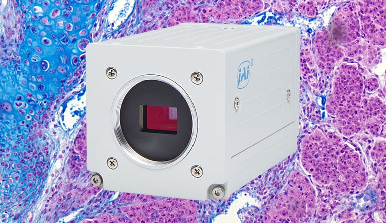
The new camera variations include both standard JAI green housings, as well as special white housings for systems intended for hospitals, or clinical/laboratory environments where white housings are often preferred.
More importantly, the new variants include some models without standard IR-cut filters. System builders can use these models to support life sciences applications where enhanced red sensitivity is required to accentuate specific dyes or stains or to differentiate blood or blood vessels from surrounding tissues. These models are also useful for certain food and industrial applications where both visible and NIR analyses are performed simultaneously. These capabilities are further bolstered by a built-in color enhancement function included as a standard feature in the Apex Series cameras.
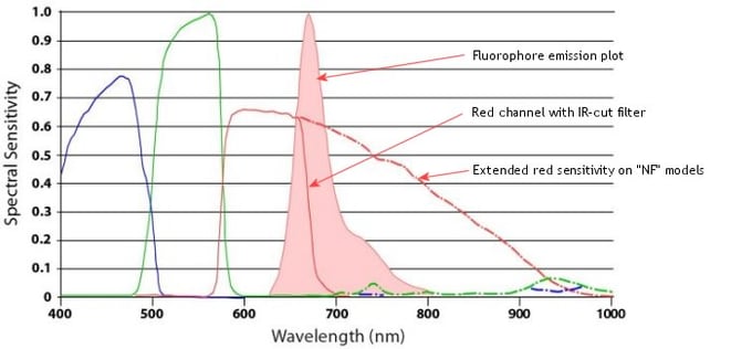
Spectral response of JAI microscopy cameras with emission plot from a common protein detection fluorophore. Note the amount of the plot that falls beyond the standard IR-cutoff point.
White Paper
If you want to learn more about the supreme color image quality provided by JAI's 3-CMOS prism based area scan cameras, you can download the free white paper: How does prism technology help to achieve superior color image quality?
Microscopy software
In conjunction with the rollout of the new models, JAI and Media Cybernetics of Rockville, MD, have jointly announced the integration of the Apex Series cameras with the popular Image-Pro image![]() analysis software platform. Image-Pro is one of the most widely-used microscopy software packages in the world, enabling users and system designers to more easily capture, process, measure, analyze and share microscopy-based images. Custom drivers allow the Apex Series cameras to seamlessly pass images to the Image-Pro software while allowing certain camera functions to be controlled from within the Image-Pro environment.
analysis software platform. Image-Pro is one of the most widely-used microscopy software packages in the world, enabling users and system designers to more easily capture, process, measure, analyze and share microscopy-based images. Custom drivers allow the Apex Series cameras to seamlessly pass images to the Image-Pro software while allowing certain camera functions to be controlled from within the Image-Pro environment.
For those who prefer an open source, non-commercial software solution, the Apex Series ![]() cameras have also been integrated with the µManager (Micro-Manager) software originally created by Vale Lab at the University of California, San Francisco and now being developed and maintained by Open Imaging, Inc. Again, device adapters have been developed to allow the JAI cameras to fully interact with the µManager software. There are no licensing fees for the software, which can be downloaded directly from the micro-manager.org website. A variety of support plans are offered by Open Imaging.
cameras have also been integrated with the µManager (Micro-Manager) software originally created by Vale Lab at the University of California, San Francisco and now being developed and maintained by Open Imaging, Inc. Again, device adapters have been developed to allow the JAI cameras to fully interact with the µManager software. There are no licensing fees for the software, which can be downloaded directly from the micro-manager.org website. A variety of support plans are offered by Open Imaging.
Applications
The combination of the new 3-CMOS color camera models and the supported software packages provides a range of new possibilities for designers and users of microscope-based systems.
With their standard C-mount lens mounts, the Apex Series cameras can be easily adapted to most brands of digital microscopes. The 3-CMOS prism technology provides much greater color differentiation than standard Bayer color cameras for brightfield or color fluorescence applications where subtle color differences are critical. Furthermore, with higher frame rates than previous generations of 3-sensor prism cameras, the AP-3200T-USB (38.3 fps) and AP-1600T-USB (78.8 fps) provide the added performance needed for live cell imaging, time-lapse microscopy, extended depth-of-field, or other color microscopy applications where real-time speeds or higher are required.
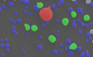
Additional applications where the color accuracy and spatial precision of the JAI 3-CMOS prism-based technology can provide advantages over traditional Bayer color cameras include wafer inspection and metal inspection systems utilizing color to classify defects; materials science including food, composites, cosmetics, and many other materials; and manufacturing systems that perform particle analysis, spray/spot analysis, or inspection of fibers or other small details.
A few specific application examples: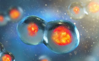
Live cell imaging - With frame rates of 38 fps or 79 fps the JAI microscopy cameras are very suitable for time-lapse microscopy in live cell imaging and in the general study of cellular dynamics.
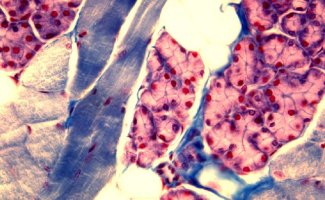
Pathology - Supreme color reproduction, high spatial resolution, and color enhancement tools make the Apex Series cameras ideal for microscopy-based systems used for tissue slice analysis, stain analysis, cell counting, cell classification and other applications in the pathology field.
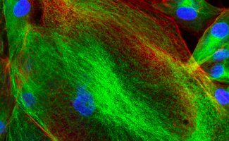
Fluorescence microscopy - Excellent color differentiation, low noise, and extended red sensitivity supports a wide range of fluorescence applications including bacteria counting, disease diagnosis, neuron signaling, and many more.
Materials science - Advanced color systems are needed for analysis of a variety of manufactured materials including coatings, cosmetics, metals, plastics, and composites.
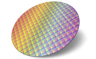
Wafer inspection - Very accurate color image data and high spatial resolution ensure precise error detection and classification of semiconductor defects in advanced wafer inspection systems.